Optic Nerve Pit – March, 2025

History:
An 11-year-old female was referred by an outside retina specialist as a second opinion for reduced visual acuity in the right eye (OD) of unknown duration. There is no prior ocular surgical history and she wore glasses for distance.
Exam:
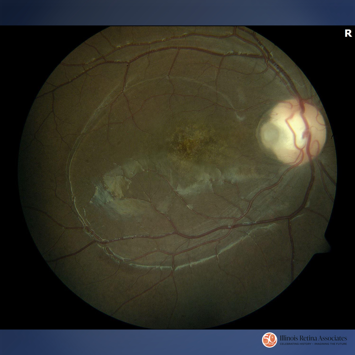
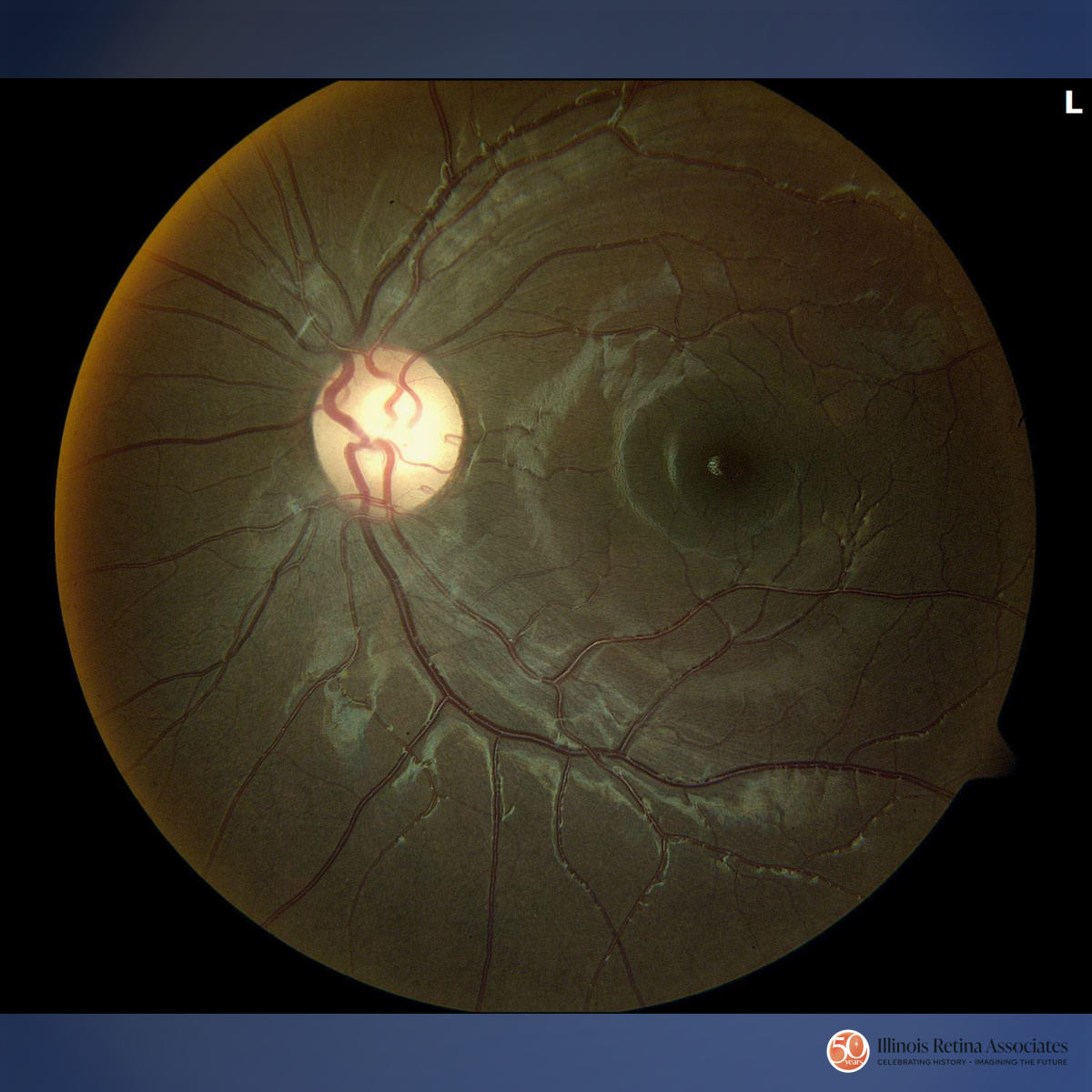
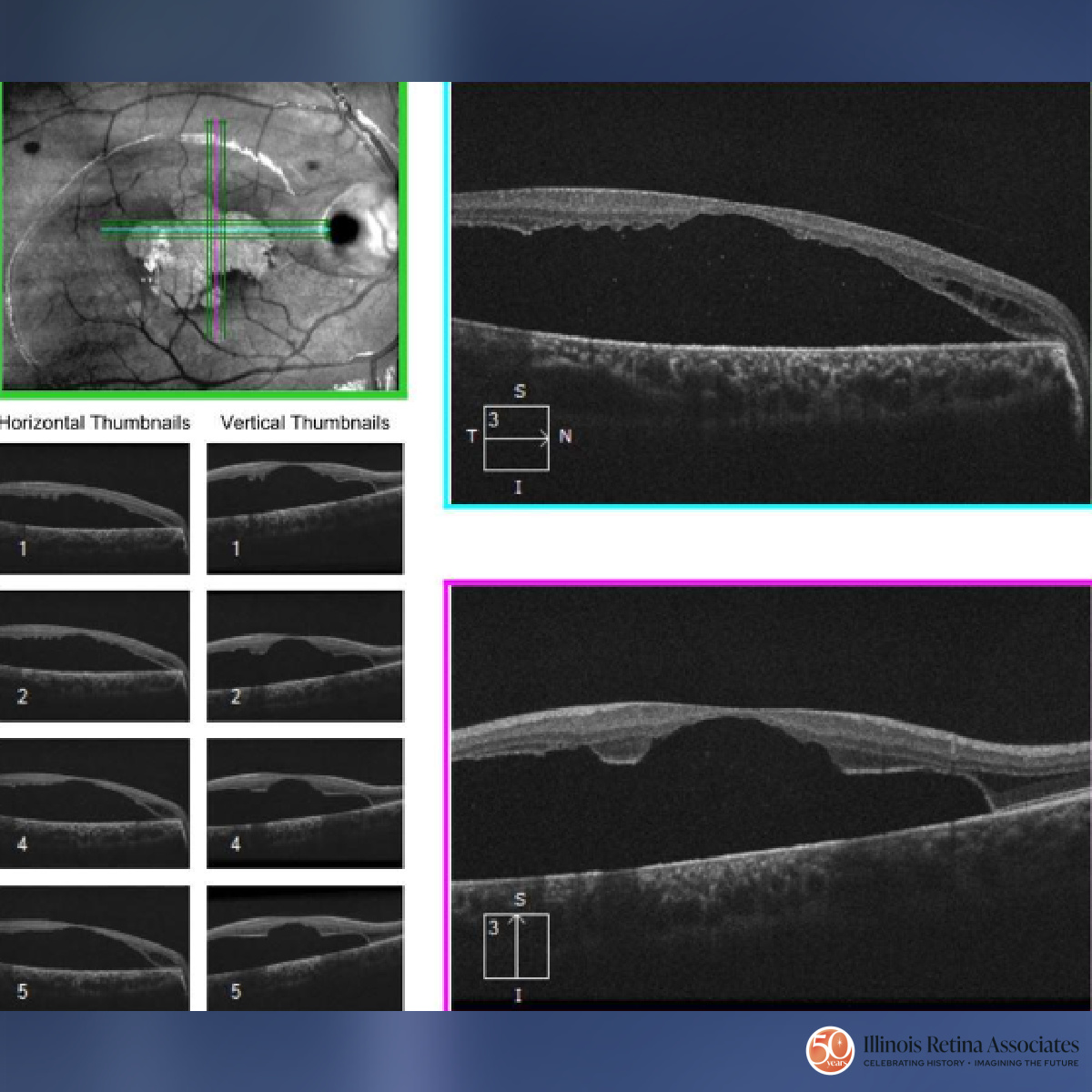
Visual acuity (VA) was 20/300 OD and 20/25 in the left eye (OS). Intraocular pressures and anterior segment exam were normal in both eyes (OU). Dilated fundus exam OD revealed a temporal optic nerve pit. Within the macula, there was subretinal fluid and retinal pigment epithelium mottling (Fig 1). Fundus exam OS was normal (Fig 2). OCT macula OD demonstrated peripapillary intraretinal fluid and foveal subretinal fluid (Fig 3).
Clinical Course:
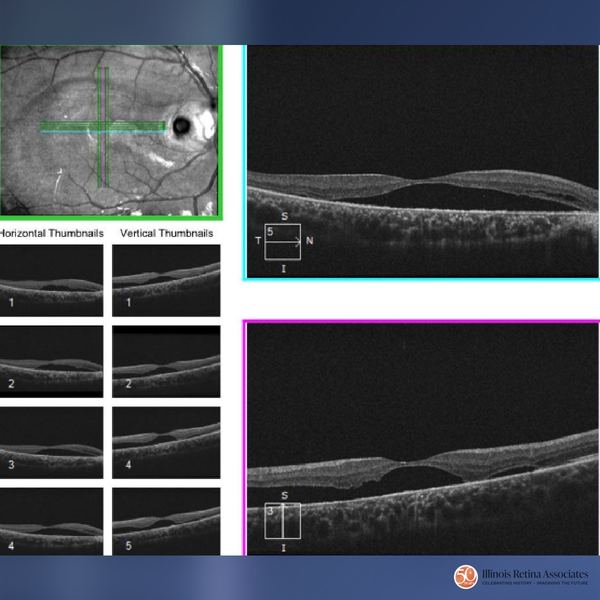
The patient underwent a pars plana vitrectomy, light endolaser to the temporal edge of the optic nerve, and gas injection of 25% SF6 gas. One month after surgery, vision improved to 20/150 with improvement of subretinal fluid on OCT (Fig 4).
Differential Diagnosis:
- Optic nerve pit
- Optic disc coloboma
- Morning glory disc anomaly
- Hypoplastic optic nerve
- Glaucoma
Discussion:
Optic Nerve Pit
Optic nerve pits are rare anomalies of the disc which results from incomplete closure of the embryonic fissure. They are usually incidental unilateral findings with pits seen in the inferotemporal disc. Visual symptoms can occur in up to 75% of patients due to maculopathy.
Maculopathy can include serous retinal detachment, cystoid macular edema, or retinoschisis. Most of these patients will progress to a VA of 20/200 or worse. The pathophysiology of serous retinal detachment is poorly understood, with sources of the fluid postulated to be from the vitreous cavity or cerebrospinal fluid from direct communication with the subarachnoid space. Typically, patients are symptomatic as they reach the third or fourth decade of life.
Management is observation unless there is macular involvement, then options include laser photocoagulation, pars plana vitrectomy with or without inverted ILM flap with gas tamponade. Pars plana vitrectomy can lead to visual improvement in over half of patients with resolution of subretinal fluid in 50-95%.
If you are looking to schedule your first consultation, please contact us today by clicking HERE and find the location that is nearest you!
References:
- Gordon R, Chatfield RK. Pits in the optic disc associated with macular degeneration. The British journal of ophthalmology. 1969;53(7):481–9.
- Chatziralli I, Theodossiadis P, Theodossiadis GP. Optic disk pit maculopathy: Current management strategies. Vol. 12, Clinical Ophthalmology. Dove Medical Press Ltd; 2018. p. 1417–22.
- Sobol WM, Blodi CF, Folk JC, et al. Long-term visual outcome in patients with optic nerve pit and serous retinal detachment of the macula. Ophthalmology 1990; 97: 1539–1542.