Laser Induced Retinal Injury – April, 2025
History:
A 20-year-old man with no significant past medical history noticed abrupt painless vision loss in his left eye (OS) while dancing at a laser-light nightclub. An ophthalmologist evaluated the patient with concern for laser-induced retinal injury. He presented to our clinic for further evaluation and treatment.
Exam:
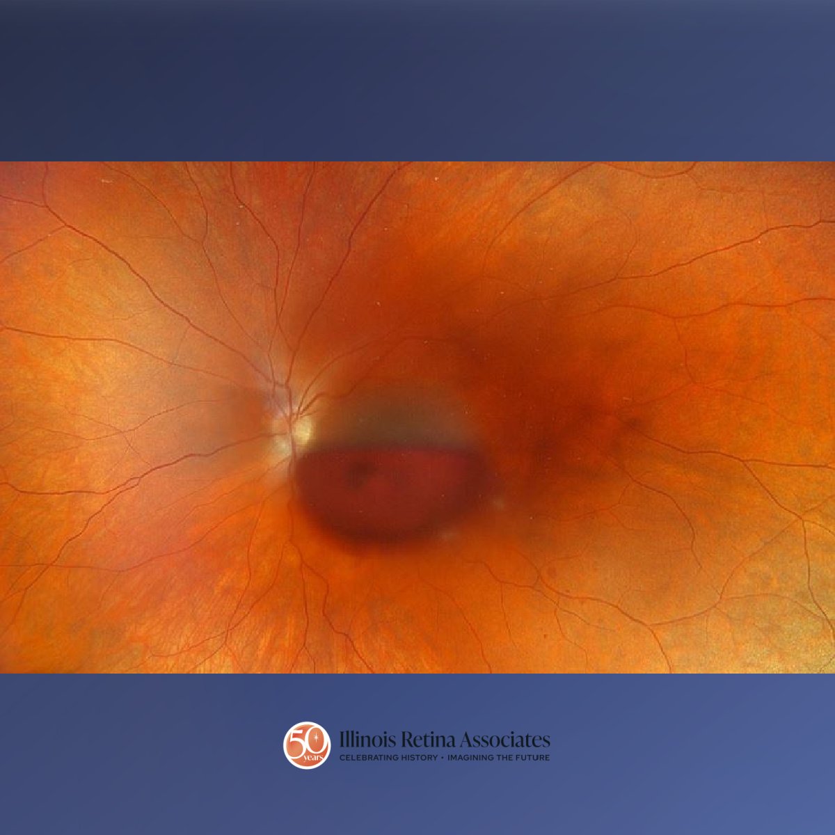
Visual acuity (VA) right eye (OD) was 20/20 with a normal exam. VA OS was count fingers with a large, center involving sub-internal limiting membrane (ILM) hemorrhage, 2 small areas of focal retinal whitening in the macula, and mild vitreous hemorrhage (Figure 1). Hematology workup was unremarkable for a clotting disorder.
Differential Diagnosis:
• Laser induced retinal injury
• Retinal vein occlusion
• Valsalva retinopathy
• Diabetic retinopathy
Discussion:
Laser Induced Retinal Injury
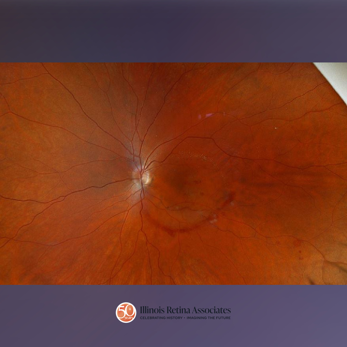
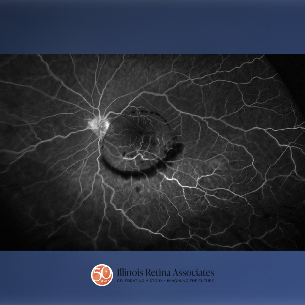
With the recent history and absence of other known medical problems, he was initially diagnosed with laser induced retinal injury. YAG laser hyaloidotomy was attempted to drain the hemorrhage, though unsuccessful. The patient elected for vitrectomy and ILM peeling. Following elevation of the posterior hyaloid, the ILM was incised with a bent 25-guage needle and the hemorrhage evacuated with a soft tip on extrusion. The ILM was peeled to clear more of the hemorrhage. The retina underneath revealed vessel tortuosity, dot blot hemorrhages, and cotton wool spots consistent with a branch retinal vein occlusion (RVO). VA was 20/50 one week following surgery (Figure 2). Ocular Coherence Tomography revealed macular thickening and fluorescein angiography showed inferior dilated and tortuous vasculature with blocking from dot hemorrhages and late staining of vessels (Figure 3). The patient subsequently discovered a remote family history of unknown clotting disorder which may have been a contributing factor to the RVO.
If you are looking to schedule your first consultation, please contact us today by clicking HERE and find the location that is nearest you!